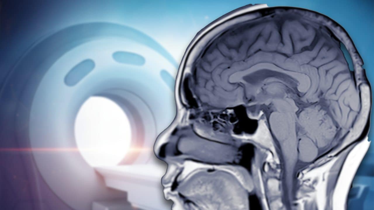
Magnetic resonance imaging – or nuclear magnetic resonance imaging – is a diagnostic technique used since the 80s to make in a non -invasive way and without the use of ionizing radiation (unlike the CT, which uses X -rays) three -dimensional images of the interior of our body. It is a technique born in the physical field in the 70s and subsequently “borrowed” by diagnostic medicine to carry out neurological, oncological, cardiological exams and so on. The only precaution: forbidden to have metal objects – including pacemakers – during the exam!
The operation of the machine for magnetic resonance imaging uses the magnetic properties of protons, particles that make up the atomic hydrogen nuclei, which are stimulated thanks to powerful magnetic fields to collect images. But how does the car do to transform the invisible signals of our atoms into such detailed images?
How the magnetic resonance imaging machine is made
The machine used for magnetic resonance imaging consists of a cylindrical tunnel in which the patient is positioned during the exam. At the center of the tunnel is a superconductor magnet that produces the magnetic field necessary for scan. This field can reach intensity of the order of 1.5-3 Tesla (some experimental machines up to 7 Tesla): so to speak, we are talking about a magnetic field tens of thousands of times more intense than the earth’s magnetic field.
Despite this, magnetic resonance imaging is a safe and non -invasive procedure. The only fundamental precaution concerns the removal of any metal object, since the magnetic field can attract ferrous materials and radio waves can heat metals on the skin.
How it works from a physical point of view
The principle of operation of magnetic resonance imaging is based on the protons of hydrogen atoms: remember that the hydrogen atom is basically a proton around which an electron “orbit”. There are two hydrogen atoms in the water (h2O) And this constitutes much of the human body: this means that the protons of hydrogen are the main responsible for the signals from which the images of magnetic resonances are produced.
A proton can in fact be thought like a small magnet, thanks to some of its quantum properties such as the intrinsic magnetic moment and its spin. In the absence of an external magnetic field, the protons are oriented completely randomly. However, if they are immersed in a strong magnetic field, here they tend to align along the direction of the field and to perform a sort of “conical movement” around this direction, known as a precession motorcycle, which is repeated with a certain frequency – called Larmor’s frequency – which depends on the intensity of the magnetic field. For example, for a magnetic field of 1.5 Tesla the Larmor frequency is around 60 Megahertz, which belongs to the radio wave band.
During the examination, the machine sends a radio wave impulse frequency equal to Larmor of the protons specific for the magnetic field produced by the machinery. At this point, the protons of the hydrogen nuclei suffer a phenomenon of resonance conceptually similar to what happens when the glasses break in response to a sound of a certain frequency: the radio waves, having the “right” frequency, transfer energy to the protons and this makes them temporarily deviate from their alignment with the main magnetic field.
When the radio impulse stops, the protons return to the state of balance by releasing excess energy. This energy is released in the form of a particular electromagnetic signal that is detected by the recipient coils of the machine and constitutes the base for the formation of the images.
How the images are produced
The signs collected are not uniforms: they vary according to the density of protons and the chemical-physical properties of the tissues, such as composition in water and lipids, molecular mobility and time that protons take to return Aligned to the main magnetic field and to lose consistency in the plan perpendicular to the magnetic field.
The coils of the machinery generate magnetic fields that gradually vary several spatial directions: this is to produce the typical rhythmic and intense sounds during the exam. These variations of the magnetic fields allow to locate the electromagnetic signals produced by protons in space, thus allowing to reconstruct three -dimensional images of the tissues.