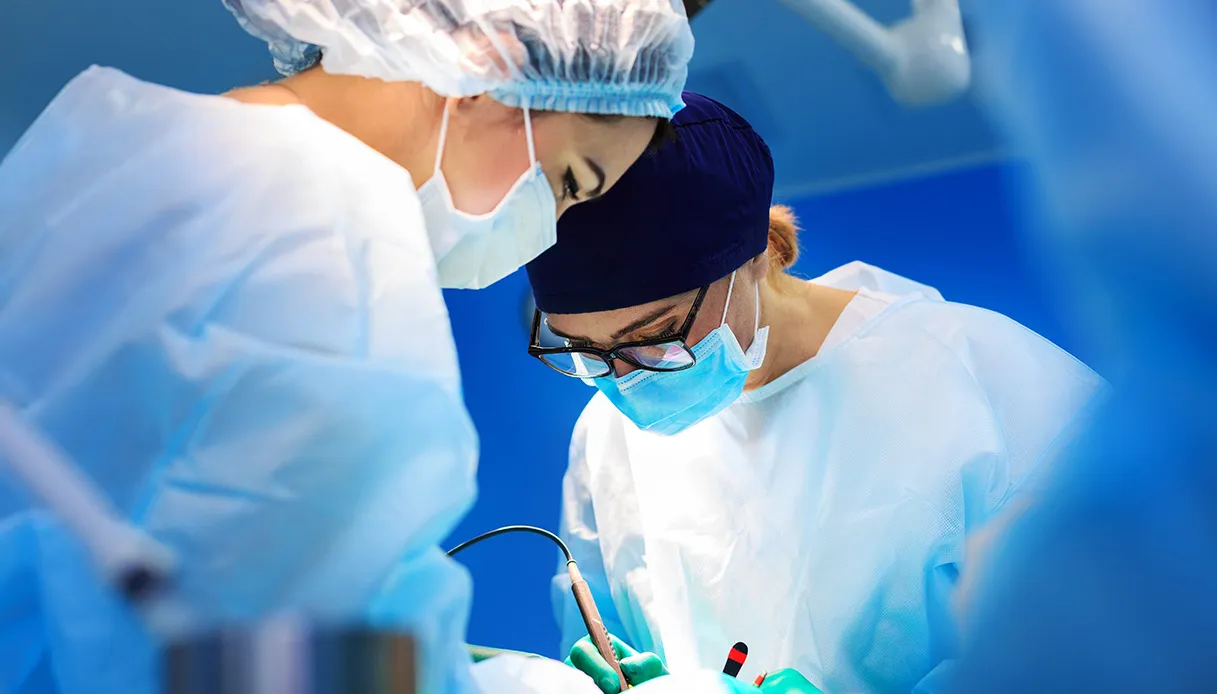
Destroy a brain tumor with laser while keeping the most delicate nervous structures of the brain under control in real time. This is the approach applied at the IRCCS Neuromed in Pozzilli, where laser thermoablation (LITT, Laser Interstitial Thermal Therapy) has been integrated with advanced anatomical-functional study and monitoring techniques of the brain.
The operation was performed on a 57-year-old patient suffering from a tumor located near the language area. The procedure took place without complications and the patient was discharged approximately 36 hours after the operation. The procedure involved a multidisciplinary team made up of neurosurgeons, neuropsychologists, neuroradiologists and anesthetists, under the guidance of Professor Sergio Paolini, head of Neurosurgery III of the IRCCS Neuromed and professor at the Sapienza University of Rome.
What the treatment involves
Laser thermoablation is a minimally invasive procedure currently performed in a few European centers and carried out at Neuromed as part of a collaboration with the Neurosurgery of the AORN Santobono Pausilipon IRCCS Pediatric Hospital in Naples, a pioneer and leader in Italy in the use of this method.
The procedure involves the introduction of an optical fiber with a diameter of less than 2 millimeters into the brain. After the positioning of the fiber, obtained robotically through a surgical microincision, the patient is transferred to MRI for the actual ablation. The laser carried by the fiber generates a controlled increase in temperature which allows the destruction of the pathological tissue. The element that characterized the procedure is the integration of the laser with the most modern brain imaging technologies.
Thanks to the use of high-field magnetic resonance imaging equipped with the most modern artificial intelligence algorithms, the team was able to follow in real time the relationship between the area subjected to the action of the laser and the eloquent brain areas, those which govern fundamental functions such as movement and language. The operation was based on an in-depth pre-operative study of the patient’s anatomy and functions integrated with the information acquired during the ablation.
“LITT is a therapeutic option applicable in selected cases of primary or metastatic brain tumors located in particularly deep or critical areas. The same method is also applicable to the removal of vascular malformations known as cavernomas. The constant challenge of neurosurgery is not only to treat the lesion but to do so while preserving the patient’s neurological functions and personality as accurately as possible”
explains Paolini.
“Modern techniques for the functional study of the brain now allow us to plan these interventions with a level of precision that was not available until a few years ago. In this case, the step forward lies in the possibility of monitoring the effects of the procedure in real time using magnetic resonance imaging.”
In particular, according to the expert,
“Minimally invasive techniques are expanding the therapeutic possibilities of modern neurosurgery. Our group has been adopting this philosophy for years in the surgery of brain and spinal tumors. However, it is important to underline that mini-invasiveness is truly feasible where it is accompanied by a reduction in surgical trauma and at the same time preserves or improves the effectiveness and safety of the procedure”.
This requirement is fully achieved by thermal ablation since the thermal necrosis of the tumor lesion is in all respects similar to surgical removal and patients can leave the hospital a few hours after the procedure.